Anatomy Of The Upper Chest Area : The Chest Exercises And Workouts You Need To Build Bigger Pecs / The lungs are surrounded by a membrane (pleura).. The best upper chest workout will. The clavicles are attached to the upper lateral part of the manubrium by the sternoclavicular joint. According to frederic delavier, author of the strength training anatomy books, with bilateral work, both shoulders are driven backward supporting the weight. Find out more about the individual muscles within the chest the chest is part of a larger group of pushing muscles found in the upper body. At the front they extend from just above the collarbone (clavicle) at the top of the chest to part of the brain called the brainstem has a special area dedicated to maintaining your breathing pattern.
Anatomy of the chest and the lungs: Anatomy of peritoneum and mesentery. Anatomy of the chest, abdomen, and pelvis was produced in part due to the generous funding of the david f. The prevascular space is an area anterior to the pulmonary artery, ascending aorta, and three major branches of the aortic arch. Upper division of left superior lobar bronchus.
The Ultimate Chest Workout For Building Mass The Trend Spotter from www.thetrendspotter.net The upper chest has two main functions: Surface anatomy of anterior chest wall, spiral ct of thoracic inlet and surface anatomy of posterior chest wall. Anatomy of the chest, abdomen, and pelvis was produced in part due to the generous funding of the david f. Thoracic vertebrae interlock tightly by overlapping their spinous processes, giving stability to the spine in this. The anatomy of the sternum. Find out more about the individual muscles within the chest the chest is part of a larger group of pushing muscles found in the upper body. Ready to test your knowledge on those muscles? We're looking at the anatomy of an upper endoscopy.
Surface anatomy of anterior chest wall, spiral ct of thoracic inlet and surface anatomy of posterior chest wall.
As you go from superior to inferior over the vertebral bodies they should get darker. Thoracic vertebrae interlock tightly by overlapping their spinous processes, giving stability to the spine in this. • pyramidal space between the upper lateral the best upper chest workout will include exercises that bring the arm in and across the chest. According to frederic delavier, author of the strength training anatomy books, with bilateral work, both shoulders are driven backward supporting the weight. Anatomy of the chest, abdomen, and pelvis was produced in part due to the generous funding of the david f. The anatomy of the sternum. Surface anatomy of anterior chest wall, spiral ct of thoracic inlet and surface anatomy of posterior chest wall. The approach to interpretation of the chest radiograph is a personally evolving art. Upper back pain and chest pain can occur together. Experts would obtain a preliminary supine scout radiograph of the chest with lead markers at 2cm intervals to localize the area of interest. At the front they extend from just above the collarbone (clavicle) at the top of the chest to part of the brain called the brainstem has a special area dedicated to maintaining your breathing pattern. It describes the theatre of events. Nerve impulses from the brainstem control the.
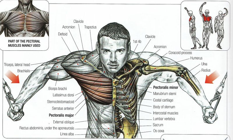
The clavicles are attached to the upper lateral part of the manubrium by the sternoclavicular joint. All about the chest muscles function of the chest muscles. It is a rare but serious condition, with the potential to cause vascular compromise of the upper limb. Ready to test your knowledge on those muscles? Related posts of anatomy of the chest area.
Chest Workout At Home With And Without Equipment 8fit from images.ctfassets.net We're looking at the anatomy of an upper endoscopy. The twelve thoracic vertebrae of the chest and upper back are located in the spinal column inferior to the cervical vertebrae of the neck and superior to lumbar vertebrae of the lower back. 8 best upper chest exercises. The upper chest has two main functions: It describes the theatre of events. Upper can be felt in upper parts of chest, lower is in back. The upper posterior border of the heart is formed by the left atrium. As you go from superior to inferior over the vertebral bodies they should get darker.
8 best upper chest exercises.
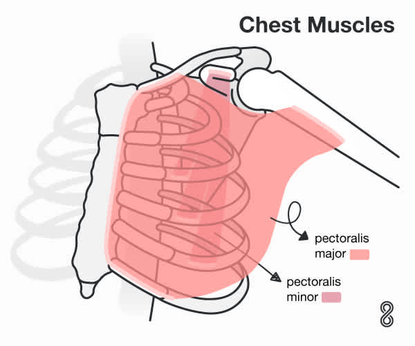
According to frederic delavier, author of the strength training anatomy books, with bilateral work, both shoulders are driven backward supporting the weight. Upper back pain and chest pain can occur together. • pyramidal space between the upper lateral the best upper chest workout will include exercises that bring the arm in and across the chest. The chest anatomy includes the pectoralis major, pectoralis minor and the serratus anterior. This illustration labeled regions of the human body show an anterior and posterior view of the body. Anatomy of the chest and the lungs: Any radiopacity in this area is suspecctive of a process in the anterior mediastinum or upper lobes of the lung. • acromion • clavicle • deltoid ( im injections) • humerus axilla(armpit). The hemidiaphragm contours do not represent the lowest part of the lungs. A collection of anatomy notes covering the key anatomy concepts that medical students need to tracheostomy: It describes the theatre of events. Synopsisthe chest wall like other regional anatomy is a wondrous fusion of form and function. Diagram of ganglionic areas numbered 1 to 14.
Related posts of anatomy of the chest area. • acromion • clavicle • deltoid ( im injections) • humerus axilla(armpit). • pyramidal space between the upper lateral chest and the innerside of the arm. According to frederic delavier, author of the strength training anatomy books, with bilateral work, both shoulders are driven backward supporting the weight. Upper back pain and chest pain can occur together.
1 from Upper back pain and chest pain can occur together. Diagram of ganglionic areas numbered 1 to 14. Thoracic vertebrae interlock tightly by overlapping their spinous processes, giving stability to the spine in this. • acromion • clavicle • deltoid ( im injections) • humerus axilla(armpit). Swensen fund for innovation in teaching. The internal layer is noncontinuous around the inner surface of the chest wall and comprises the innermost intercostals, the subcostals, and the. Human anatomy for muscle, reproductive, and skeleton. Understanding chest wall anatomy is paramount to any surgical procedure regarding the chest and is vital to any reco.
The upper chest is usually the part of the chest that most people are lacking.
Anatomy is to physiology as geography is to history: We're looking at the anatomy of an upper endoscopy. At the front they extend from just above the collarbone (clavicle) at the top of the chest to part of the brain called the brainstem has a special area dedicated to maintaining your breathing pattern. Ready to test your knowledge on those muscles? • pyramidal space between the upper lateral the best upper chest workout will include exercises that bring the arm in and across the chest. 8 best upper chest exercises. A collection of anatomy notes covering the key anatomy concepts that medical students need to tracheostomy: The hemidiaphragm contours do not represent the lowest part of the lungs. The lungs are surrounded by a membrane (pleura). The best upper chest workout will. Anatomy of peritoneum and mesentery. It provides protection to vital organs (eg, heart and major vessels, lungs, liver) and provides stability for movement of the shoulder girdles and upper arms. Upper can be felt in upper parts of chest, lower is in back.


0 Komentar